LUMC uses artificial intelligence to calculate lung damage in coronavirus patients
With the aid of artificial intelligence (AI), care professionals at the LUMC (Leiden University Medical Center) are able to calculate quickly and accurately whether a coronavirus patient has suffered serious lung damage. They do this by putting a CT scan through the AI software of the CAD4COVID-CT program.
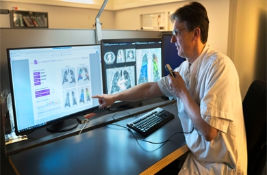
Coronavirus patients admitted to the hospital are generally given a CT scan to determine the nature and gravity of a lung infection. The assessment of these scans has now been automated, thanks to the CAD4COVID-CT AI software. The software determines how serious the coronavirus infection is based on the percentage of damaged lung tissue found. This saves precious time for doctors and radiologists.
The software was developed by Thirona and is used at the LUMC in the Philips Intellispace AI Workflow Suite. The application of AI belongs to the Clinical Artificial Intelligence Implementation and Research Lab (CAIRELab), a knowledge and expertise centre for all AI work at the LUMC. The Radiology Department is one of the hospital’s partners and a frontrunner in the application of AI. The data from the quantitative image analysis enables care professionals to make a quick and accurate diagnosis of the gravity of the disease, facilitates a prediction of its progression in patients.

‘A radiologist’s human intelligence is well able to recognise patterns,’ says Hildo Lambe, Professor of Radiology at the LUMC. ‘But the human brain has more difficulty with extremely accurate quantification and calculations of deviating areas. The support of AI makes this extreme accuracy possible and saves time, so we can diagnose COVID-19 more quickly and accurately, and monitor more efficiently whether the treatment is taking effect.’
Radiologist always observer
After every calculation, a report in PDF is saved to the system. These reports are all viewed and assessed by a radiologist at the LUMC. ‘The report contains a quantification of how much damage there has been to lung tissue in the various lung lobes,’ Lambe explains. ‘The system also creates an overlay image, so that the radiologist can see the data on which the algorithm based its choice.’
Big step for AI in healthcare
CAD4COVID-CT was developed by Thirona in response to the coronavirus pandemic. ‘We wanted to contribute’, Mark van Grinsven, head of product development at Thirona, tells us. ‘We’re happy about the collaboration with Philips, which allows us to bring AI to the foreground of healthcare and offers users of the LUMC a fully integrated workflow. We think this is a wonderful initial step towards automatic analysis of medical images with AI.’
The LUMC is the first hospital in the world to introduce Intellispace AI Workflow Suite, which offers care providers the possibility of integrating AI applications into the workflow.
