Dangerous microbes in lower level safety lab? A new technique could make it possible
Researchers need to work in specialized environments when they work with dangerous bacteria and viruses. These microbes spread easily, so only in labs with a high biosafety levels they can be studied. Unfortunately, to look at the microbes properly, expensive microscopes are needed that are not always present in these extra safe labs. New research shows a new way of how dangerous viruses and bacteria can be handled.

‘With our research, we use a very large microscope to look at very small things. In this way, we get a detailed understanding of how microbes work,’ says Jamie Depelteau, a freshly graduated PhD of the Briegel lab and first author of the article published in Communications Biology. ‘We look at the microbes, which are made up of proteins, to determine how they interact with the environment.’
To do this type of research, researchers often apply a chemical fixation. This inactivates or kills the microbes, which makes dangerous variants safe to work with. Yet, a major downside for this technique is that the chemicals alter the structure of the bacteria or virus. An unwelcome side effect when studying the microbes’ surface.
UV as solution
A solution came up after a lot of brainstorming in the Briegel lab, and with collaborators in the Jensen Lab at Caltech: UVC irradiation. Depelteau: ‘We constructed a simple light box by which we could treat the samples with ultraviolet C light. UVC targets the DNA of viruses and bacteria leading to inactivation within seconds or minutes.’ But the question remained whether this UVC treatment also damaged the microbe’s protein structure?
Glass-like ice for perfect vision
To test the effect of the UVC irradiation technique, Depelteau et al. used the cryo-electron microscopy. 'With cryo-EM, we surround the samples with water and cool them very quickly to -184 degrees Celsius. And because it happens so quickly, the water doesn’t crystallize. Instead, it turns into a glass-like state, which we can see through perfectly. That allows electrons to travel through, with which we can zoom in extensively on the microbes.’ Once frozen, images were collected using the top-of-the-line microscropes at the Netherlands Centre for Electron Nanoscopy (NeCEN) located at Leiden University.

From micrometers to sub-nanometer
First, Depelteau et al. examined UVC-treated bacteria. ‘We looked at the treated cells and did not see any visible damage. So we zoomed in on the bacterial nose, the chemotaxis array, which they use to sense its environment and move to nutritious places, for example. Also in this case, everything was still clear. And more importantly: it looked the same as when we compared it to images in the untreated sample. UVC irradiation did not obviously alter the bacteria’s nose,’ Depelteau proclaims happily.

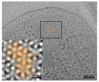
Since cryo-EM allows researchers to zoom in even further, they also looked at a virus. ‘Which worked very well too!’ And even when examining the coat of the virus, Depelteau et al. could accurately discern different sections. ‘We have gone across scales, from micrometers with the bacteria, to nanometers with the virus and you could even say sub-nanometer with the protein, and all appears well.’
A new tool in the toolbox for studying dangerous microbes
Being able to study proteins safely and accurately this way can make a big difference, according to Depelteau. ‘All beings are basically made up of proteins. It is important to see what proteins look like and how they react in certain environments. Especially for dangerous viruses and bacteria. If researchers use this technique, there is the potential to study these pathogens safely and accurately. Using UVC irradiation could become an important tool in the protein-studying toolbox.’
Read the complete article in Communication Biology
Depelteau, J.S., Renault, L., Althof, N. et al. UVC inactivation of pathogenic samples suitable for cryo-EM analysis. Commun Biol 5, 29 (2022). https://doi.org/10.1038/s42003-021-02962-w
