
Can we live longer? Leiden physicist makes discovery in protective layer in genes
With the aid of physics and a minuscule magnet, researchers have discovered a new structure of telomeric DNA. Telomeres are sometimes seen as the key to living longer. They protect genes from damage but get a bit shorter each time a cell divides. If they become too short, the cell dies. The new discovery will help us understand ageing and disease.
Physics is not the first scientific discipline that springs to mind at the mention of DNA. But John van Noort from the Leiden Institute of Physics (LION) is one of the scientists who found the new DNA structure. A biophysicist, he uses methods from physics for biological experiments. This also caught the attention of biologists from Nanyan Technological University in Singapore. They asked him to help study the DNA structure of telomeres. They have published the results in Nature.
String of beads
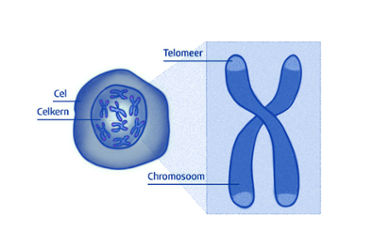
In every cell of our bodies are chromosomes that carry genes that determine our characteristics (what we look like, for instance). At the ends of these chromosomes are telomeres, which protect the chromosomes from damage. They’re a bit like aglets, the plastic tips at the end of a shoelace.
The DNA between the telomeres is two metres long, so it has to be folded to fit in a cell. This is achieved by wrapping the DNA is wrapped around packages of proteins; together, the DNA and proteins are called a nucleosome. These are arranged into something similar to a string of beads, with a nucleosome, a piece of free (or unbound) DNA, a nucleosome and so on.
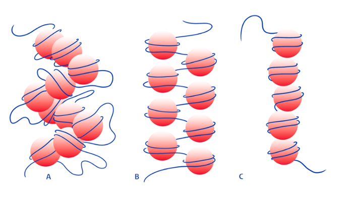
This string of beads then folds up even more. How it does so depends on the length of the DNA between the nucleosomes, the beads on the string. Two structures that occur after folding were already known. In one of them, two adjacent beads stick together and free DNA hangs in between (fig. 2A). If the piece of DNA between the beads is shorter, the adjacent beads do not manage to stick together. Then two stacks form alongside each other (fig. 2B).
In their study Van Noort and colleagues found another telomere structure. Here the nucleosomes are much closer together, so there is no longer any free DNA between the beads. This ultimately creates one big helix, or spiral, of DNA (fig. 2C).
Magnet
The new structure was discovered with a combination of electron microscopy and molecular force spectroscopy. The latter technique comes from Van Noort’s lab. Here one end of the DNA is attached to a glass slide and a tiny magnetic ball is stuck to the other. A set of strong magnets above this ball then pull the string of pearls apart. By measuring the amount of force needed to pull the beads apart one by one, you find out more about how the string is folded. The researchers in Singapore then used an electron microscope to get a better picture of the structure.
Building blocks
Structure, says Van Noort, is ‘the holy grail of molecular biology.’ If we know the structure of the molecules, this will give us more insight into how genes are switched on and off and how enzymes in cells deal with telomeres: how they repair and copy DNA, for example. The discovery of the new telomeric structure will improve our understanding of the building blocks in the body. And that in turn will ultimately help us study ageing and diseases such as cancer and develop drugs to fight them.
Text: Dagmar Aarts
Images: Fien Leeflang