

From nanoscale to whole organism: at the Cell Observatory, researchers study life in detail
About forty microscopes, various laboratories, and some 15,000 zebrafish: that’s Sylvia le Dévédec's workplace. She is one of the managers of the Leiden Cell Observatory, a unique facility accessible to all researchers.

Le Dévédec walks down the long corridor, enthusiastically pointing out various labs where cultures are developed and rooms with microscopes. At the end is the zebrafish enclosure. She frequently encounters students and colleagues with whom she strikes up a conversation. ‘You meet people from all kinds of fields here, that’s what makes this work so valuable.’
Together with biologist Joost Willemse and chemist Amit Cherian, the LACDR researcher manages the microscopy section of the Cell Observatory: a shared facility where anyone in need of a microscope can go. Because, as Le Dévédec says, it makes no sense for different departments to buy the same expensive microscope; it is better to share. ‘This way, we curate a varied collection that allows us to answer more research questions.’
‘Life is incredibly beautiful’
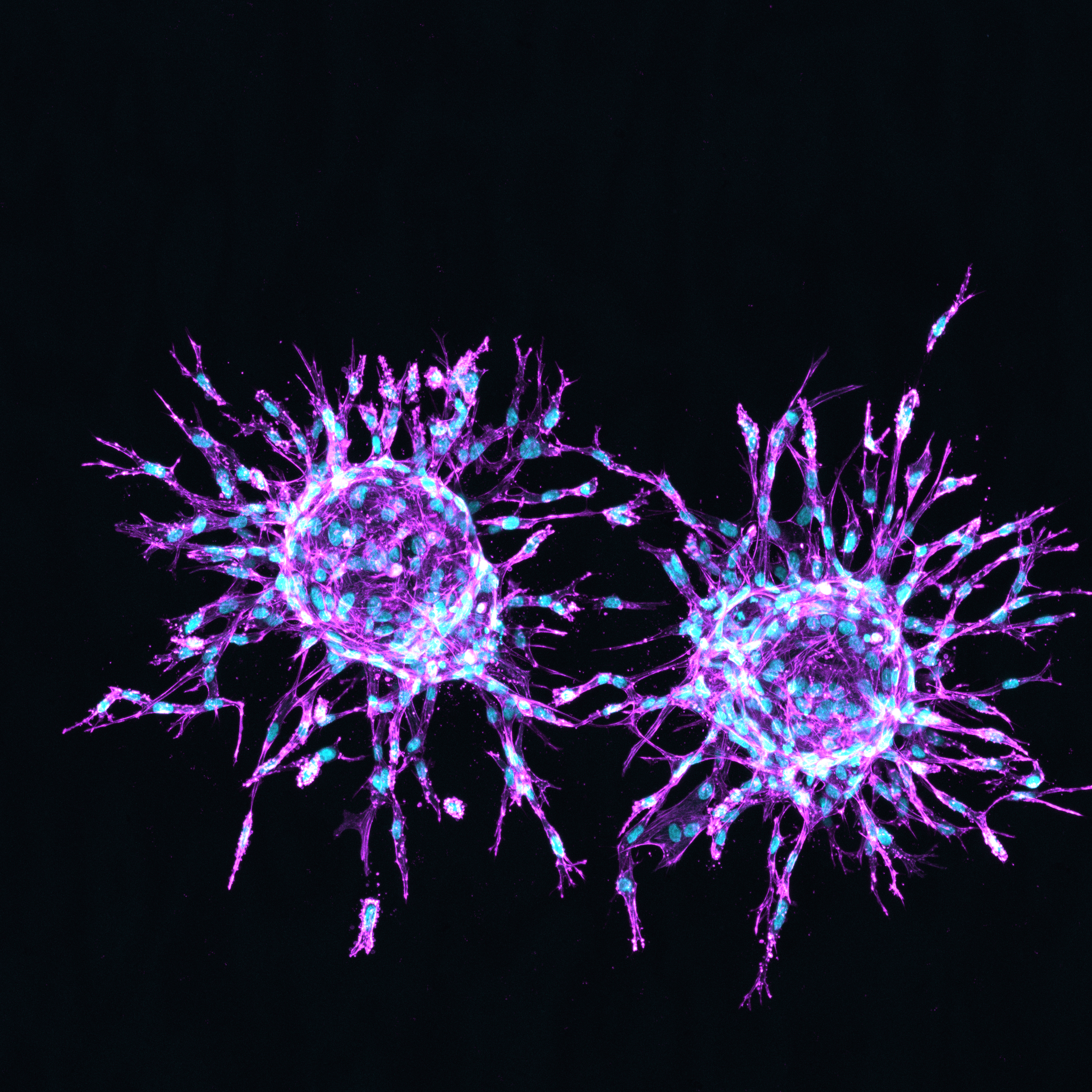
In the Cell Observatory’s annual photo competition, researchers compete for the title of the most beautiful picture. ‘I’ve had many wow moments behind the microscope myself,’ says Le Dévédec. ‘You see living cells moving with your own eyes; that is so incredibly beautiful.’
-

First prize of the 2025 photo competition: Max FernKorn with 'Everything is connected' -
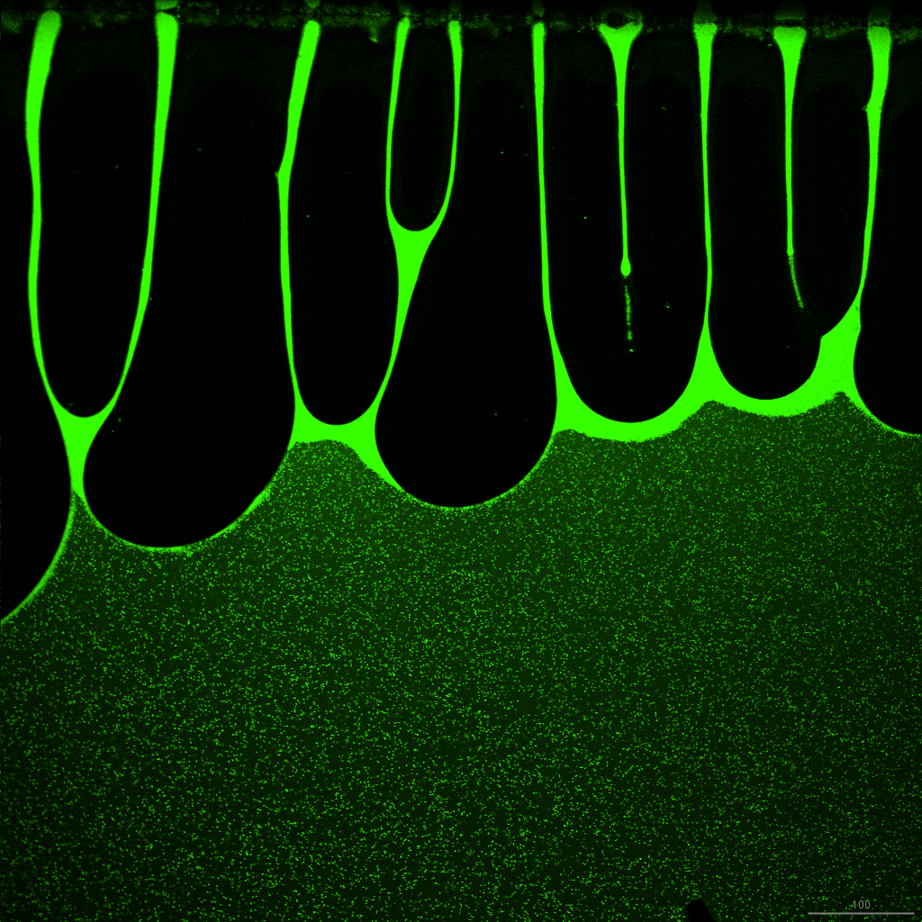
Second prize: Alex Versluis with 'The touch of creation' -
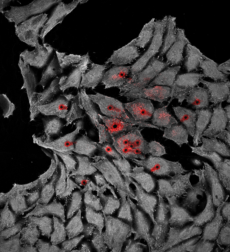
Third prize: David Norte with 'Sticky Fingers' -
Fourth prize: Don Schilder with 'Wounds of light' -
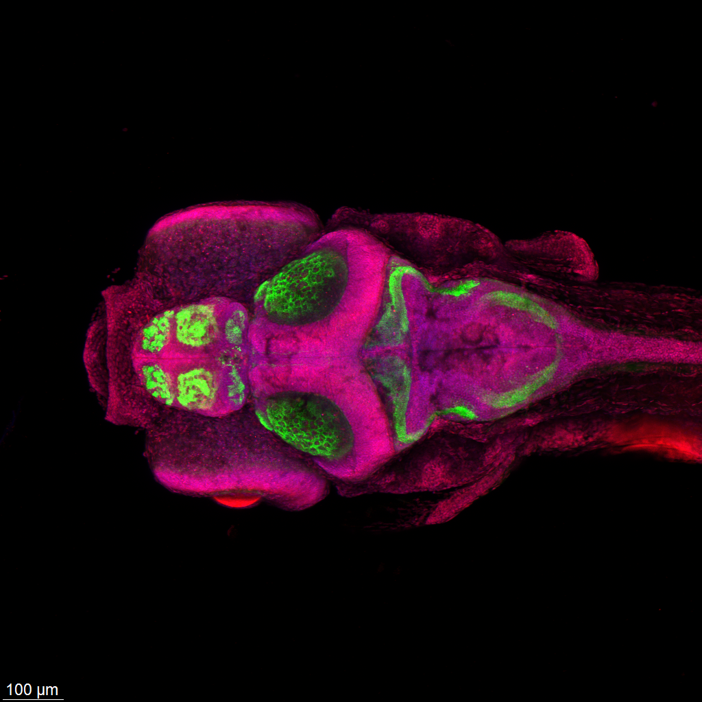
Fifth prize: Joaquin Abugattas-Nuñez Del Prado with 'Neural Architecture in Bloom'
A closer look at metastasising cancer cells
Le Dévédec’s research focuses on breast cancer in the hope of eventually inhibiting metastases. How do cancer cells spread through the body? That is where microscopy comes into play. ‘I try to understand the role of certain genes in cell migration,’ she explains. ‘In a 96 well plate (a plastic plate the size of a hand with 96 wells), we can test a different condition in each well. If you see the phenotype—the physical characteristics, that is—change under the microscope, then you know that the gene played a role.’

A huge leap in innovation in just one generation
Things were very different during her PhD, Le Dévédec recalls. ‘I could only investigate one condition at a time because I had to manually track a few cells under the microscope all day long. Now, we can photograph up to a million cells fully automatically in a single weekend.’
She shows a microscope with a whole tower of 96 well plates. These are placed in a room with precisely the right temperature and humidity to keep the cells alive. A robotic arm picks up the plates one by one and places them under the microscope, which subsequently photographs each well. Le Dévédec: ‘So much is possible these days.’

Microscopy brings science to life
As manager of the facility, she primarily supports the users. From advice on which microscope to use to applying for funding to purchase new equipment. She would also like to collaborate more with the business community at the Leiden Bio Science Park: ‘For small companies, it is often too expensive to purchase such microscopes. I would love to offer them the opportunity to use our facilities and learn from our experience.’
Whether it concerns her own research, supervising PhD candidates, or managing the Cell Observatory: her passion is palpable. ‘Microscopy brings science to life,’ says Le Dévédec. ‘It’s not just numbers, you really see what happens inside living cells. That is why I enjoy going to work every day.’